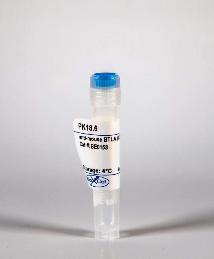
InVivoMAb anti-mouse BTLA (CD272)
| Clone | PK18.6 | ||||||||||||
|---|---|---|---|---|---|---|---|---|---|---|---|---|---|
| Catalog # | BE0153 | ||||||||||||
| Category | InVivoMab Antibodies | ||||||||||||
| Price |
|
The PK18.6 monoclonal antibody reacts with mouse B- and T-lymphocyte attenuator (BTLA) also known as CD272. BTLA is an Ig superfamily member which is expressed on B cells, T cells, macrophages, dendritic cells, NK cells, and NKT cells. Like PD-1 and CTLA-4, BTLA interacts with a B7 homolog, B7-H4. However, unlike PD-1 and CTLA-4, BTLA displays T cell inhibition via interaction with tumor necrosis family receptors, not just the B7 family of cell surface receptors. BTLA is a ligand for herpes virus entry mediator (HVEM). BTLA-HVEM complexes have been shown to negatively regulate T cell immune responses.
| Isotype | Rat IgG1, κ |
| Recommended Isotype Control(s) | InVivoMAb rat IgG1 isotype control, anti-horseradish peroxidase |
| Recommended Dilution Buffer | InVivoPure™ pH 7.0 Dilution Buffer |
| Immunogen | Mouse BTLA external domain-human IgG fusion protein |
| Reported Applications |
|
| Formulation |
|
| Endotoxin |
|
| Purity |
|
| Sterility | 0.2 μM filtered |
| Production | Purified from tissue culture supernatant in an animal free facility |
| Purification | Protein G |
| RRID | AB_10949465 |
| Molecular Weight | 150 kDa |
| Storage | The antibody solution should be stored at the stock concentration at 4°C. Do not freeze. |
nVivoMAb anti-mouse BTLA (CD272) (Clone: PK18.6)
Krieg, C., et al. (2005). "Functional analysis of B and T lymphocyte attenuator engagement on CD4+ and CD8+ T cells." J Immunol 175(10): 6420-6427. PubMed
T cell activation can be profoundly altered by coinhibitory and costimulatory molecules. B and T lymphocyte attenuator (BTLA) is a recently identified inhibitory Ig superfamily cell surface protein found on lymphocytes and APC. In this study we analyze the effects of an agonistic anti-BTLA mAb, PK18, on TCR-mediated T cell activation. Unlike many other allele-specific anti-BTLA mAb we have generated, PK18 inhibits anti-CD3-mediated CD4+ T cell proliferation. This inhibition is not dependent on regulatory T cells, nor does the Ab induce apoptosis. Inhibition of T cell proliferation correlates with a profound reduction in IL-2 secretion, although this is not the sole cause of the block of cell proliferation. In contrast, PK18 has no effect on induction of the early activation marker CD69. PK18 also significantly inhibits, but does not ablate, IL-2 secretion in the presence of costimulation as well as reduces T cell proliferation under limiting conditions of activation in the presence of costimulation. Similarly, PK18 inhibits Ag-specific T cell responses in culture. Interestingly, PK18 is capable of delivering an inhibitory signal as late as 16 h after the initiation of T cell activation. CD8+ T cells are significantly less sensitive to the inhibitory effects of PK18. Overall, BTLA adds to the growing list of cell surface proteins that are potential targets to down-modulate T cell function.
Han, P., et al. (2004). "An inhibitory Ig superfamily protein expressed by lymphocytes and APCs is also an early marker of thymocyte positive selection." J Immunol 172(10): 5931-5939. PubMed
Positive selection of developing thymocytes is associated with changes in cell function, at least in part caused by alterations in expression of cell surface proteins. Surprisingly, however, few such proteins have been identified. We have analyzed the pattern of gene expression during the early stages of murine thymocyte differentiation. These studies led to identification of a cell surface protein that is a useful marker of positive selection and is a likely regulator of mature lymphocyte and APC function. The protein is a member of the Ig superfamily and contains conserved tyrosine-based signaling motifs. The gene encoding this protein was independently isolated recently and termed B and T lymphocyte attenuator (Btla). We describe in this study anti-BTLA mAbs that demonstrate that the protein is expressed in the bone marrow and thymus on developing B and T cells, respectively. BTLA is also expressed by all mature lymphocytes, splenic macrophages, and mature, but not immature bone marrow-derived dendritic cells. Although mice deficient in BTLA do not show lymphocyte developmental defects, T cells from these animals are hyperresponsive to anti-CD3 Ab stimulation. Conversely, anti-BTLA Ab can inhibit T cell activation. These results implicate BTLA as a negative regulator of the activation and/or function of various hemopoietic cell types.


