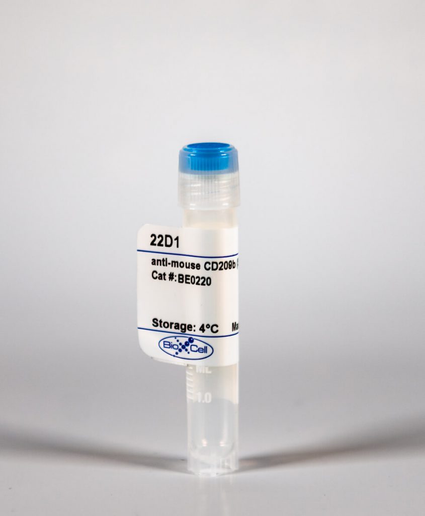
InVivoMab anti-mouse CD209b (SIGN-R1)
| Clone | 22D1 | ||||||||||||
|---|---|---|---|---|---|---|---|---|---|---|---|---|---|
| Catalog # | BE0220 | ||||||||||||
| Category | InVivoMab Antibodies | ||||||||||||
| Price |
|
The 22D1 monoclonal antibody reacts with mouse CD209b also known as SIGN-R1. CD209b is a 37 kDa type II transmembrane C-type lectin receptor. CD209b is expressed on the surface of splenic marginal zone and lymph node medullary macrophages and is commonly used as a marker for these cells. The CD209b protein is involved in the innate immune response, it binds to and initiates uptake of various microorganisms by recognizing high-mannose-containing glycoproteins on their envelopes. The 22D1 antibody has been reported to block CD209b in vivo.'
| Isotype | Armenian Hamster IgG, κ |
| Recommended Isotype Control(s) | InVivoMAb polyclonal Armenian hamster IgG(BE0091) |
| Recommended InVivoPure Dilution Buffer | InVivoPure pH 7.0 Dilution Buffer(IP0070) |
| Immunogen | C-terminal peptide of mouse SIGN-R1 |
| Reported Applications |
|
| Endotoxin |
|
| Purity |
|
| Formulation |
|
| Sterility | 0.2 μM filtered |
| Production | Purified from tissue culture supernatant in an animal free facility |
| Purification | Protein G |
| Storage | The antibody solution should be stored at the stock concentration at 4°C. Do not freeze. |
| RRID | AB_2687704 |
| Molecular Weight | 150 kDa |
InVivoMAb anti-mouse CD209b (SIGN-R1) (Clone: 22D1)
Zinselmeyer, B. H., et al. (2013). "PD-1 promotes immune exhaustion by inducing antiviral T cell motility paralysis." J Exp Med 210(4): 757-774. PubMed
Immune responses to persistent viral infections and cancer often fail because of intense regulation of antigen-specific T cells-a process referred to as immune exhaustion. The mechanisms that underlie the induction of exhaustion are not completely understood. To gain novel insights into this process, we simultaneously examined the dynamics of virus-specific CD8(+) and CD4(+) T cells in the living spleen by two-photon microscopy (TPM) during the establishment of an acute or persistent viral infection. We demonstrate that immune exhaustion during viral persistence maps anatomically to the splenic marginal zone/red pulp and is defined by prolonged motility paralysis of virus-specific CD8(+) and CD4(+) T cells. Unexpectedly, therapeutic blockade of PD-1-PD-L1 restored CD8(+) T cell motility within 30 min, despite the presence of high viral loads. This result was supported by planar bilayer data showing that PD-L1 localizes to the central supramolecular activation cluster, decreases antiviral CD8(+) T cell motility, and promotes stable immunological synapse formation. Restoration of T cell motility in vivo was followed by recovery of cell signaling and effector functions, which gave rise to a fatal disease mediated by IFN-gamma. We conclude that motility paralysis is a manifestation of immune exhaustion induced by PD-1 that prevents antiviral CD8(+) T cells from performing their effector functions and subjects them to prolonged states of negative immune regulation.
Moens, L., et al. (2007). "Specific intracellular adhesion molecule-grabbing nonintegrin R1 is not involved in the murine antibody response to pneumococcal polysaccharides." Infect Immun 75(12): 5748-5752. PubMed
Streptococcus pneumoniae is a microorganism that frequently causes serious infections in children, the elderly, and immunocompromised patients. We studied whether the specific intracellular adhesion molecule-grabbing nonintegrin R1 (Sign-R1) receptor, involved in the uptake of capsular polysaccharides (caps-PS) by antigen-presenting cells, is necessary for the antibody response to pneumococcal caps-PS and phosphorylcholine (PC). The antibody response to caps-PS and PC was evaluated after vaccination with soluble caps-PS (Pneumovax) and after vaccination with heat-killed S. pneumoniae. The role of Sign-R1 was investigated by using Sign-R1 knockout mice and anti-Sign-R1 monoclonal antibodies. The immunoglobulin M (IgM) and IgG antibody response to PC and caps-PS (serotypes 3 and 14) was not affected by anti-Sign-R1 monoclonal antibodies. The IgM antibody response in Sign-R1 knockout mice was comparable to the antibody response in wild-type mice. The IgG antibody response to serotype 3, but not to serotype 14, tended to be lower in Sign-R1 knockout mice compared to wild-type mice. In conclusion, we found that Sign-R1 is not involved in the IgM antibody production to PC and caps-PS serotype 3 or 14 and the IgG immune response to PC and caps-PS serotype 14. There is no direct relation between capture and uptake of caps-PS serotype 14 by Sign-R1 and the initiation of the anti-caps-PS antibody production in mice.
Kang, Y. S., et al. (2004). "The C-type lectin SIGN-R1 mediates uptake of the capsular polysaccharide of Streptococcus pneumoniae in the marginal zone of mouse spleen." Proc Natl Acad Sci U S A 101(1): 215-220. PubMed
SIGN-R1, a recently discovered C-type lectin expressed at high levels on macrophages within the marginal zone of the spleen, mediates the uptake of dextran polysaccharides by these phagocytes. We now find that encapsulated Streptococcus pneumoniae are rapidly cleared by these macrophages from the bloodstream, and that capture also takes place when different cell lines express SIGN-R1 after transfection. To assess the role of the capsular polysaccharide of S. pneumoniae (CPS) in the interaction of SIGN-R1 with pneumococci, we first studied binding and uptake of serotype 14 CPS in transfected cells. Binding was observed and was of a much higher avidity (3000-fold) for CPS 14 than dextran. The CPSs from four different serotypes were also cleared by marginal zone macrophages in vivo. To establish a role for SIGN-R1 in this uptake, we selectively down-regulated expression of the lectin by pretreatment of the mice with SIGN-R1 antibodies, including a newly generated hamster monoclonal called 22D1. For several days after this transient knockout, the marginal zone macrophages were unable to take up either CPSs or dextrans. Therefore, marginal zone macrophages in mice have a receptor that interacts with capsular pneumococcal polysaccharides, setting the stage for further studies of the functional consequences of this interaction.


