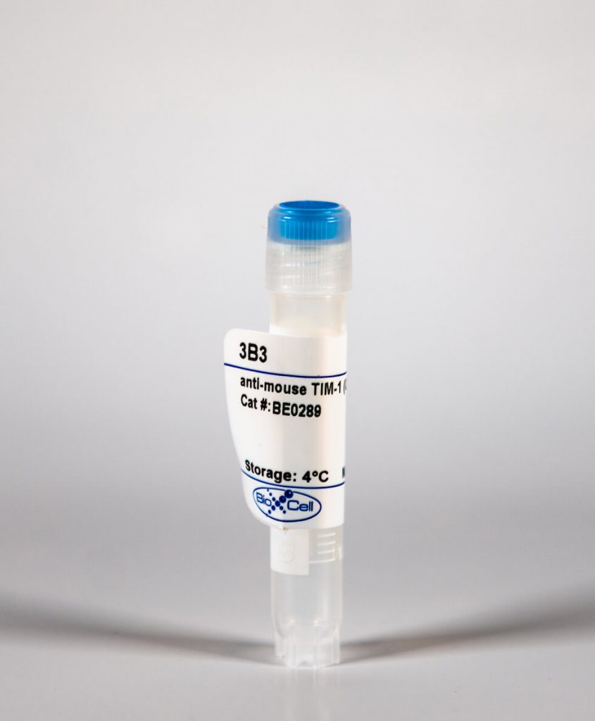
InVivoMab anti-mouse TIM-1 (CD365)
| Clone | 3B3 | ||||||||||||
|---|---|---|---|---|---|---|---|---|---|---|---|---|---|
| Catalog # | BE0289 | ||||||||||||
| Category | InVivoMab Antibodies | ||||||||||||
| Price |
|
The 3B3 monoclonal antibody reacts with mouse T cell immunoglobulin and mucin domain 1 (TIM-1) also known as CD365. TIM-1 is a type I cell-surface glycoprotein and member of the Ig superfamily. TIM-1 is preferentially expressed on TH2 cells and has been identified as a stimulatory molecule for T cell activation. The TIM gene family, plays critical roles in regulating the immune response to viral infection. TIM-1 is also involved in allergic responses, asthma, and transplant tolerance. The 3B3 antibody is an agonistic antibody and has been shown to activate TIM-1 in vivo and in vitro resulting in the hyperproliferation of T cells and induction of proinflammatory cytokine production.
| Isotype | Rat IgG2a, κ |
| Recommended Isotype Control(s) | InVivoMAb rat IgG2a isotype control, anti-trinitrophenol |
| Recommended Dilution Buffer | InVivoPure™ pH 7.0 Dilution Buffer |
| Immunogen | Mouse TIM-1 (signal and IgV domains)/mouse IgG2a Fc fusion protein |
| Reported Applications |
|
| Formulation |
|
| Endotoxin |
|
| Purity |
|
| Sterility | 0.2 μM filtered |
| Production | Purified from tissue culture supernatant in an animal free facility |
| Purification | Protein G |
| RRID | AB_2687812 |
| Molecular Weight | 150 kDa |
| Storage | The antibody solution should be stored at the stock concentration at 4°C. Do not freeze. |
INVIVOMAB ANTI-MOUSE TIM-1 (CD365) (CLONE: 3B3)
Lee, H. H., et al. (2010). “Apoptotic cells activate NKT cells through T cell Ig-like mucin-like-1 resulting in airway hyperreactivity.” J Immunol 185(9): 5225-5235. PubMed
T cell Ig-like mucin-like-1 (TIM-1) is an important asthma susceptibility gene, but the immunological mechanisms by which TIM-1 functions remain uncertain. TIM-1 is also a receptor for phosphatidylserine (PtdSer), an important marker of cells undergoing programmed cell death, or apoptosis. We now demonstrate that NKT cells constitutively express TIM-1 and become activated by apoptotic cells expressing PtdSer. TIM-1 recognition of PtdSer induced NKT cell activation, proliferation, and cytokine production. Moreover, the induction of apoptosis in airway epithelial cells activated pulmonary NKT cells and unexpectedly resulted in airway hyperreactivity, a cardinal feature of asthma, in an NKT cell-dependent and TIM-1-dependent fashion. These results suggest that TIM-1 serves as a pattern recognition receptor on NKT cells that senses PtdSer on apoptotic cells as a damage-associated molecular pattern. Furthermore, these results provide evidence for a novel innate pathway that results in airway hyperreactivity and may help to explain how TIM-1 and NKT cells regulate asthma.
Mariat, C., et al. (2009). “Tim-1 signaling substitutes for conventional signal 1 and requires costimulation to induce T cell proliferation.” J Immunol 182(3): 1379-1385. PubMed
Differentiation and clonal expansion of Ag-activated naive T cells play a pivotal role in the adaptive immune response. T cell Ig mucin (Tim) proteins influence the activation and differentiation of T cells. Tim-3 and Tim-2 clearly regulate Th1 and Th2 responses, respectively, but the precise influence of Tim-1 on T cell activation remains to be determined. We now show that Tim-1 stimulation in vivo and in vitro induces polyclonal activation of T cells despite absence of a conventional TCR-dependent signal 1. In this model, Tim-1-induced proliferation is dependent on strong signal 2 costimulation provided by mature dendritic cells. Ligation of Tim-1 upon CD4(+) T cells with an agonist anti-Tim-1 mAb elicits a rise in free cytosolic calcium, calcineurin-dependent nuclear translocation of NF-AT, and transcription of IL-2. Because Tim-4, the Tim-1 ligand, is expressed by mature dendritic cells, we propose that interaction between Tim-1(+) T cells and Tim-4(+) dendritic cells might ensure optimal stimulation of T cells, when TCR-derived signals originating within an inflamed environment are weak or waning.
Degauque, N., et al. (2008). “Immunostimulatory Tim-1-specific antibody deprograms Tregs and prevents transplant tolerance in mice.” J Clin Invest 118(2): 735-741. PubMed
T cell Ig mucin (Tim) molecules modulate CD4(+) T cell responses. In keeping with the view that Tim-1 generates a stimulatory signal for CD4(+) T cell activation, we hypothesized that an agonist Tim-1-specific mAb would intensify the CD4(+) T cell-dependant allograft response. Unexpectedly, we determined that a particular Tim-1-specific mAb exerted reciprocal effects upon the commitment of alloactivated T cells to regulatory and effector phenotypes. Commitment to the Th1 and Th17 phenotypes was fostered, whereas commitment to the Treg phenotype was hindered. Moreover, ligation of Tim-1 in vitro effectively deprogrammed Tregs and thus produced Tregs unable to control T cell responses. Overall, the effects of the agonist Tim-1-specific mAb on the allograft response stemmed from enhanced expansion and survival of T effector cells; a capacity to deprogram natural Tregs; and inhibition of the conversion of naive CD4(+) T cells into Tregs. The reciprocal effects of agonist Tim-1-specific mAbs upon effector T cells and Tregs serve to prevent allogeneic transplant tolerance.
Xiao, S., et al. (2007). “Differential engagement of Tim-1 during activation can positively or negatively costimulate T cell expansion and effector function.” J Exp Med 204(7): 1691-1702. PubMed
It has been suggested that T cell immunoglobulin mucin (Tim)-1 expressed on T cells serves to positively costimulate T cell responses. However, crosslinking of Tim-1 by its ligand Tim-4 resulted in either activation or inhibition of T cell responses, thus raising the issue of whether Tim-1 can have a dual function as a costimulator. To resolve this issue, we tested a series of monoclonal antibodies specific for Tim-1 and identified two antibodies that showed opposite functional effects. One anti-Tim-1 antibody increased the frequency of antigen-specific T cells, the production of the proinflammatory cytokines IFN-gamma and IL-17, and the severity of experimental autoimmune encephalomyelitis. In contrast, another anti-Tim-1 antibody inhibited the generation of antigen-specific T cells, production of IFN-gamma and IL-17, and development of autoimmunity, and it caused a strong Th2 response. Both antibodies bound to closely related epitopes in the IgV domain of the Tim-1 molecule, but the activating antibody had an avidity for Tim-1 that was 17 times higher than the inhibitory antibody. Although both anti-Tim-1 antibodies induced CD3 capping, only the activating antibody caused strong cytoskeletal reorganization and motility. These data indicate that Tim-1 regulates T cell responses and that Tim-1 engagement can alter T cell function depending on the affinity/avidity with which it is engaged.
Umetsu, S. E., et al. (2005). “TIM-1 induces T cell activation and inhibits the development of peripheral tolerance.” Nat Immunol 6(5): 447-454. PubMed
We have examined the function of TIM-1, encoded by a gene identified as an ‘atopy susceptibility gene’ (Havcr1*), and demonstrate here that TIM-1 is a molecule that costimulates T cell activation. TIM-1 was expressed on CD4(+) T cells after activation and its expression was sustained preferentially in T helper type 2 (T(H)2) but not T(H)1 cells. In vitro stimulation of CD4(+) T cells with a TIM-1-specific monoclonal antibody and T cell receptor ligation enhanced T cell proliferation; in T(H)2 cells, such costimulation greatly enhanced synthesis of interleukin 4 but not interferon-gamma. In vivo, the use of antibody to TIM-1 plus antigen substantially increased production of both interleukin 4 and interferon-gamma in unpolarized T cells, prevented the development of respiratory tolerance, and increased pulmonary inflammation. Our studies suggest that immunotherapies that regulate TIM-1 function may downmodulate allergic inflammatory diseases.


